MMC Instruments
-
DMi 1, Leica DMi8 inverted fluorescence tissue scanning, deconvolution microscope
-
DMi 2, Leica DMi8 inverted fluorescence tissue scanning microscope
-
Live Imaging System Zeiss Axio Observer.Z1 with Apotome 2
-
Light Sheet and confocal microscope
-
Zeiss LSM 710 Confocal. 6 laser lines, 3 detectors
-
Leica Stellaris 8 Confocal. White Light Laser (multiplex imaging), Lightning deconvolution, lifetime imaging, live imaging
-
Seahorse metabolic analysis
Overview of Services
The Medicine Microscopy Core (MMC) provides a user-friendly, cutting-edge fluorescence microscopy imaging environment for the Columbia Medical Center in Upper Manhattan. We also provide image analysis tools, such as Leica Aivia 3D machine learning software and an Acquifer HIVE computer server and workstation. We are happy to collaborate with your imaging and image analysis projects and can provide software, custom scripting, and custom acquistion pipelines.
Click on the tabs (or icon lines) above to explore the microscopes.
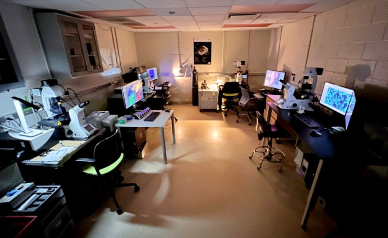
Primary imaging room. Black Building, room 828.
Medicine Microscopy Core (MMC)
iLab Link for ReservationsContact (replace © and •): jwm2175©cumc•columbia•edu
News
2026-04-28DMi station 2 has been repaired and is working normally. The filter turret motor was replaced by R. Brideau.
2026-04-08DMi station 1 has been repaired thanks to excellent work of Leica Service rep, Rob Brideau. The system is working normally. See me if you need special objective lenses for your work.
2026-03-31Unfortunately, both DMi station 1 and DMi station 2 are not working properly. DMi station 2 can still be used to image 1 channel at a time. See me for instructions.
... see all News
Staff
John W Murray, PhD
Imaging Facility Director
Assistant Professor
Columbia University
Department of Medicine
Columbia Center for Human Development (CCHD)
jwm2175©cumc•columbia•edu
Office phone: 212•305•4130
Coordinates
William Black Building
(google maps link)
8th Floor, Room 828, 832, and VP&S 8-508B
Office: room 801F
650 West 168th Street
New York, NY 10032
Hours
Hours: 24/7
Staffed: M-F, 9am-5pm


© 2026 Columbia Medicine Microscopy Core (MMC)